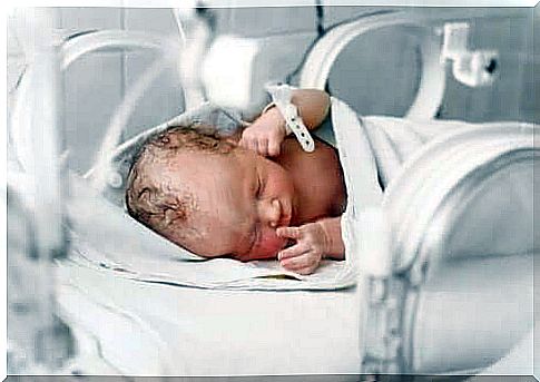
Gastroschisis In Infants
The prenatal disease gastroschisis is also known as the abdominal cleft. Surgical intervention is usually carried out after the birth, otherwise this malformation could be fatal.
The gastroschisis is one of the most common congenital malformations, which is, however, largely unknown. A defect in the abdominal wall usually occurs spontaneously to the right of the navel as long as the child is still in the womb. The result is a prolapse of intestinal loops, which means that the child’s abdominal contents (intestines or stomach) get into the amniotic sac and swim in the amniotic fluid.
This can cause damage to the bowel, which can shorten, swell, or become tangled. Once the child is born, surgery is required to bring the abdominal contents back into place and close the abdominal wall. Despite this surgical intervention , gastroschisis can lead to problems with nutrition, digestion and nutrient absorption.
Gastroschisis: causes and risk factors
In some cases, gastroschisis is due to genetic changes. A combination of genetic factors and certain circumstances during pregnancy can also cause it. The following factors increase the risk of the baby suffering from gastroschisis:
- Pregnancy at a very young age: Adolescent mothers are at greater risk that the unborn child will suffer from gastroschisis.
- Tobacco and alcohol use: Babies of women who smoke or consume alcohol are also more at risk of having a cleft abdomen.
Gastroschisis: diagnosis and treatment
The diagnosis of gastroschisis can be made before birth or immediately after birth. If a prenatal diagnosis with fine ultrasound is performed during pregnancy, this disease can be detected. In addition, there are specific examinations for diagnosis.
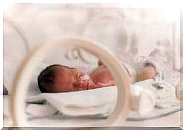
Treatment of an abdominal cleft is fundamental because this malformation can be fatal. The doctor performs the surgical procedure as soon as possible after the birth of the child so that the organs can develop properly and lie protected in the abdomen.
If the defect is small, a single surgical procedure is enough. However, if the abdominal gap is larger and several organs emerge from the abdominal cavity, the operation is carried out in several steps. After the surgery, the infant spends some time in the neonatal intensive care unit.
Risks of Surgical Intervention
The general risks of local anesthesia and surgery are usually:
- Allergic reactions to medication
- Difficulty breathing
- Infection and bleeding
The specific risks of the procedure to correct gastroschisis are:
- Difficulty breathing: These can occur when the child’s abdomen is smaller than normal. The newborn may need artificial respiration a few days or weeks after surgery.
- Swelling of the tissue covering the abdominal wall
- Injury to the organs
- Occasional paralysis of the small intestine
- Hernia on the abdominal wall
Complementary treatments
In addition to the surgical procedure , the following therapies are often necessary:
- Nasogastric nutrition to keep the stomach clear.
- Intravenous hydration and nutrient supply
- oxygen
- Antibiotics to prevent infections
- Painkiller
After the operation, nutrition is provided via a nasal tube as soon as the child’s bowel function begins again. Oral nutrition must be introduced slowly and gradually. The doctor usually discharges the child from the hospital 15 to 25 days after the operation.
Prevention against gastroschisis
Preventive measures consist of proper prenatal care and healthy habits during pregnancy. Regular check-ups are also important to prevent complications. Once the diagnosis has been made, specialized care and proper monitoring are essential.
In this case, ultrasound examinations play an important role. A weekly ultrasound scan is recommended from the 30th week of pregnancy.
Most babies born with gastroschisis are light. In addition, between 10 and 20 percent suffer from bowel malformations. The extent of these malformations largely determines the prognosis that the doctor will make.
In the last few years the chances of survival have improved a lot. This is due to early diagnosis and also to the use of specific protocols for prenatal monitoring of the child. Of course, appropriate care measures are also essential for the development of the baby.